Saturday 1 July 2017

Xrayted - Xray Answers


Time for answers!  Each answer corresponds to the slide number!


1.  Normal
2.  Right lower/mid zone pneumonia - patch consolidation
3.  Pulmonary oedema - a classic exam question is list the 6 xray findings of heart failure/pulmonary oedema

A - Alveolar Oedema (Haziness around the hlium looks like bat wings)
B - Kerly B lines
C - Cardiomegaly
D - Dilated prominant upper lobe vessels
E - Pleural Effusions
F - Fluid in the horizontal fissure (right lung)


4.  Left hilar mass - well circumscribed coin shape lesion likely lung cancer
5.  Pneumoperitoneum
6.  Large right sided pleural effusion
7.  Large well circumscribed lesion in the right midzone likely lung carcinoma
8.  Tension pneumothorax on the right side - note the tracheal and cardiac deviation AWAY from the lesion
9.  Multiple pulmonary metastasis - no they are NOT cannonball metastasis from a renal tumour, remember to check the two important areas, behind the heart and the apices this is a left apical primary - Pancoast Tumour
10. Aspiration pneumonia with a oesophageal stent - bilateral lower zone patchy consolidation R>L and a  oesophageal stent seen through the mediastinum
11. Bilateral pneumotharacies
12. Bilateral pleural effusions with a R sided tension pneumothorax - note the tracheal and cardiac deviation
13. A very large heart!  - This is a pericardial effusion
14. A white out in the left lung, the tracheal deviation away from the lesion makes this a large pleural effusion
15. My favourite X-ray! You can see three causes of air on this x-ray - this is Tuberculosis

  • Pneumothorax - Right upper zone
  • Cavitating lesions - throughout the chest, most easily seen in the right upper zone
  • Air Bronchogram - This is a small air like tree seen in the right mid zone, this is present in pneumonia differentiating this from lobar collapse
Congratulations if you got them all right!

Thursday 29 June 2017

Xrayted - Chest Xray Interpretation


Today we're looking at basic CXR interpretation, a topic that I have seen come up at all stages of undergraduate assessment.  Basic radiological anatomy is essential for 1st and 2nd years students and by the end of medical school all students should have developed a system to look at chest xrays.  In this blog post I will quickly refresh anatomy and walk you through a common system used to interpret a chest xray.  To finish there is a quiz with 15 of my favourite patients i've seen (progressive difficulty!)

I'm sure everyone knows basic chest anatomy.  Ive seen the corresponding picture appear in junior year OSCEs with students having to label the diagram.

Film Description

In an OSCE setting examiners will be expecting you to comment on the details of the film itself.  You have to state the following:

1) Patient Name    2) Area of Xray   3) Orientation of the film AP/lateral/PA  (it should state on the Xray)

Next is the quality of the image looking at the following:

1) Rotation - Each head of the clavicle should be equidistant from the spinous process.
2) Penetration - The intervertebral discs should be visible behind the heart.
3) Inspiration - 9 posterior ribs should be seen before the diaphram

A great summary of these with questions can be found HERE

Image interpretation

Some people use the ABCDE approach but I find it awkward and unnatural in order.  I would advocate a visual approach, starting at the top and moving down through the middle and then back up through the lungs:

  • Trachea - Is it central? It should overlie the vertebrae
  • Mediastinum - Is it enlarged? If >3cm  it is
  • Heart - Is there cardiomegaly? It should be less than half of the thorax ?Can I see the heart borders?
  • Diaphram - Are they elevated?  Any gas below them?
  • Costophrenic angles - Are they visable?  Obscuration may suggest a small pleural effusion
  • Lung Fields - Any abnormalities, opacification, lung markings
  • Bones - Any rib fractures?  How do the shoulders look?
  • Special Areas - Apices and behind the heart
Simple!  Follow those steps and you'll have no problem at examination.  Now put them to the test with the below quiz and practice going through the quality of the film spheel!  The link to the answers is below, but try to come to a definite answer and description for each xray before you look!

ANSWERS

Friday 8 February 2013

LIVER let die - Alcoholic Liver Disease and LFTs


We have a guest author this week, a Mr Philip Nelson from the University of Glasgow who has been working with the medical education department for the past 5 weeks.

 Scots have an unenviable reputation for alcohol consumption. Unfortunately this means that there are a lot of admissions to hospital due to alcohol abuse. It seems as though it is going to be a big problem in the future as compared with 30 years ago there are 4 times as many people dying of alcoholic liver disease(ALD) now.

Last week we had a tutorial on ALD, talking a bit about the functions of the liver and what to look for in LFTs. In this post we will go over ALD and have a few questions at the end you can mull over. You can view the presentation here.


The liver is a complicated organ, so it's helpful to simplify its functions into three main groups:

  • Storage - glycogen, vitamins A, D, B12, K, iron, copper
  • Metabolism - drugs, carbohydrates (gluconeogenesis, glycogenolysis), lipids, proteins 
  • Production - bile, albumin, clotting factors, hormones (inslulin like growth factor 1, thrombopoeitin)
With that in mind we will focus on how alcohol effects the liver. In chronic alcohol abuse the liver is damaged and some of these functions become impaired. There are three main pathologies that constitute ALD: 

  • Steatosis: Fatty liver is a reversible condition that occurs in nearly all people who are chronic alcohol abusers. Excess fatty acids accumulate in hepatocytes. Ususally asymptomatic. 
  • Hepatitis: Again reversible, affecting about 1 in 4 alcoholics. Hepatocyte inflammation and necrosis. Signs and symptoms include jaundice, weight loss, tender hepatomegaly, fevers.
  • Cirrhosis: Usually irreversible, affecting 10-20% of alcoholics. The liver becomes inflamed, fibrosed and undergoes micronodular regeneration.  

Patients who have cirrhosis will exhibit some of the signs of chronic liver disease, including:
- Hepatomegaly +/- Splenomegaly
- Jaundice, pruritis 
- Gynaecomastia
- Spider naevi
- Palmar erythema
- Finger Clubbing 
- Dupytrens contracture
- Asterixis
- Ascites
- Encephalopathy


When you're assessing a patient with ALD, its important to take a thorough history and examination to get an idea of the effects of the disease on the individual. Blood tests are also an important part of the assessment. The main ones to look in ALD at are: 

  • Albumin, INR - Synthetic function of the liver. Albumin decreases and INR increases in cirrhosis.
  • AST/ALT - Hepatic enzymes. Both raised, if ratio AST:ALT = >2:1 it is due to alcohol
  • ALP, GGT - Cholestatic enzymes. ALP mildly raised, GGT significantly raised.
  • Bilirubin - Conjugated by liver. Raised in cirrhosis, higher in acute hepatitis. 


Three complications of ALD that patients may present with are ascites, encephalopathy and varices. We talked about these in the presentation and the links provide another good introduction to each topic.These complications all have an effect on the prognosis of the disease, which can be estimated using the Child-Pugh score.

Measure 1 point 2 points 3 points
Total bilirubin, μmol/l <34 34-50 >50
Serum albumin, g/l >35 28-35 <28
PT INR <1.7 1.71-2.30 > 2.30
Ascites None Mild Moderate to Severe
Hepatic encephalopathy None Grade I-II (or suppressed with medication) Grade III-IV (or refractory)

Points Class One year survival Two year survival
5-6 A 100% 85%
7-9 B 81% 57%
10-15 C 45% 35%

Here are some sample LFTs for you to think about. Try to come up with differential diagnoses for each. The information on the slides might give you some help.  


Friday 11 January 2013

Danger! - Blood Gas Interpretation

After a long Christmas break I'm back to teaching and what a better way to start the year with some blood gas interpretation.

The first thing to think about before doing the gas is "will a venous sample suffice?".  This is often over looked due to habit or requests from a old school consultant.  The H+, Bicarbonate and Lactate are accepted to be the same between arterial and venous samples and the correlation with pCO2 for hypercarbia is regularly debated.  The pO2 is obviously not present in venous samples and the sample should be compared with the patients current oxygen saturation.  A good summary article of these issues can be found here.

There are 5 key questions to ask when interpreting an ABG.


1. What is the oxygenation like?  Is there respiratory failure?

Type 1 - pO2 <8    pCO2<6
Type 2 - pO2 <8    pCO2 >6

2.  Look at the H+, is there acidosis (H+ >45) or alkalosis (H+<45)?

3.  Look at the CO2, is it raised (respiratory acidosis) or lowered (respiratory alkalosis)?   Does this fit?

4.  Look at the Bicarb, is it raised (metabolic alkalosis) or lowered (metabolic acidosis)?  Does this fit?

5.  Is there any compensation? Either Full, Partial or None


Remember there only 4 broad answers to blood gas interpretation and you should know a handful of causes for each.


Respiratory Acidosis - These people have a high CO2 and are hypoventilating.  Using a top down approach the causes are:

Respiratory centre - Drugs i.e. opoids
Peripheral nerves - Guillain-Barré syndrome
Neuromuscular junction - Myaesthenia gravis
Chest Wall - Obesity, severe kyphoscoliosis
Airways - COPD, Severe asthma

Respiratory Alkalosis - These people are the opposite and are hyperventilating.  People hyperventilate commonly due to anxiety and pain, however, more serious conditions such as P.E. and Subarachnoid Haemorrhage need to be remembered

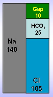
Metabolic Acidosis - These people have extra acid or have lost base.  You can calculate their anion gap to see if there are extra acids by :

(Na+ + K+) - (Cl- - HCO3-).  
The normal range is 12 - 18. 
(The reason it is not 0 is due to the weak acid affects of albumin and lactate)


If it is raised then it is due to one of the following: MUDPILES


Methanol intoxication
Uremia
Diabetic or alcoholic ketoacidosis
Propolyene Glycol
Isoniazid
Lactic acid
Ethylene glycol intoxication
Salicylate intoxication


If the gap is normal then it is likely due to Renal tubular acidosis, Diarrhea or Gastrointestinal fistula


Metabolic Alkalosis - This is more likely from the loss of acid rather than gaining base as even with infusing bicarbonate the pH barely moves.  Acid is lost through vomiting or through the kidney with diuretics or Conn's syndrome.

It is possible to have a mix of two i.e. respiratory and metabolic acidosis by combining two diagnosis' from above.  I appreciate that some people out there may prefer using the strong ion difference rather than the traditional Henderson Hasselbalch approach outlined above.  For undergraduates the traditional method is more than adequate and exams will feature this approach.  For those keen for a deeper understanding see Scott Weingarts brilliant acid base series here,

Here are 5 examples and I want you to work them through using the 5 questions and tell me the acid base disturbance and a possible cause.


1
2
3
4
5
H+  
(35-45)
62
29
27
84
103
pCO2
(4.6- 6)
10.3
2.7
5.8
2.5
7.8
pO2
(10.5 - 13)
6.5
18.1
11.4
16.3
8.8
Bic
(22-26)
32
25
35
9
6


Scroll down for the answers.


1.Type 2 Respiratory Failure with a partialy compensated respiratory acidosis COPD.  2. Uncompensated Respiratory Alkalosis Anxiety attack.  3.  Uncompensated compensated metabolic alkalosis  Vomiting.  4.  Partially compensated metabolic acidosis DKA.  5. Mixed respiratory and metabolic acidosis Obese Sepsis


Friday 30 November 2012

DKA - It does exactly what it says on the tin

Being Scottish and working in Accident and Emergency in the west of Scotland I probably see more diabetic patients than most.  A high proportion of the attendances are due to DKA.  Until today I wasn't sure why our incidence is so high.  The patient we saw today had had 14 episodes of DKA since diagnosis 2 years ago each one related to drinking excessively and forgetting to take his insulin.  He understood the reason and had been educated numerous times but drinking with his pals was too appealing to him.

Each week we start with a review of the physiology as being the basis for understanding the pathology and this week is no exception.

Exocrine Functions
Pancreatic Acini
Trypsin, chemotrypsin, lipase and amylase    
Signals from gastrin, cholecystokinin and secretin – stomach and duodenum

Endocrine
Islets of Langerhans
Alpha cells producing glucagon (15–20% of total islet cells)  Beta cells producing insulin (65–80%)
Delta cells producing somatostatin (3–10%)


This simple diagram shows glucose homeostasis

Now DKA is important and a heavily examined topic because of its incidence and high mortality.  Unbelievably pre-1920s and the use of insulin the mortality was close to 100%.  This has fallen with modern medicine to 0.7%.I think the key point of DKA is that it occurs due to a LACK OF INSULIN rather than a high blood sugar and we are treating the lack of insulin rather that the high glucose.  Without insulin the cells lack their GLUT transporters and as a result are paradoxically glucose deplete.  As a result the following cascade occurs:Lack of Insulin
  1. Reduced cellular glucose
  2. Increased glycogenolysis and gluconeogenesis
  3. Adipose and muscle metabolism causing ketone formation
  4. Glucosuria causes polyuria
  5. Ketouria and electrolyte losses
  6. Bicarbonate decreases due to acidotic ketones
  7. Respiratory compensation with tachypnoea lowers pCO2
  8. Decompensation

Diagnosis is simple, you need the three letters from the title:

D - Blood sugar >11 or known diabetic
K - Ketones +2 in the urine or >3mmol in the blood 
A - Acidotic Bic <18 or H+ >45

CAUSES

  • 1st presentation Diabetes type I
  • Not taking insulin due to poor education/poor compliance (weight issues in vain females/males)
  • Intercurrent Illness
  • Stroke or MI


MANAGEMENT 

Management should take place in a HDU setting using an ABCDE approach.

The 3 pillars of management are:
       
Insulin is given to drive cells and turn off gluconeogenesis and glycogenolysis - not to lower the blood sugar number!  The amount given in relation to the sugar is minor the key is to get some in quickly.
 

Human Insulin – Actrapid no loading dose needed
 
BM> 14 insulin at 6units/h
9-14, insulin at 3 units/h
<9, insulin at 1 unit/h
  

Fluids are to replace those lost via polyuria and increased insensible losses 

0.9% Saline

1L/hour for 2 hours
500ml/hour for 4 hours
250ml/hour for 4 hours 
 
When the blood sugar drops below 14 change the fluids to dextrose as usually more insulin will be required to turn off the gluconeogenesis and glycogenolysis than normal blood sugars allow.
  

Potassium is given even when the the levels are normal as from the management of hyperkalaemia we know insulin drives potassium into cells.